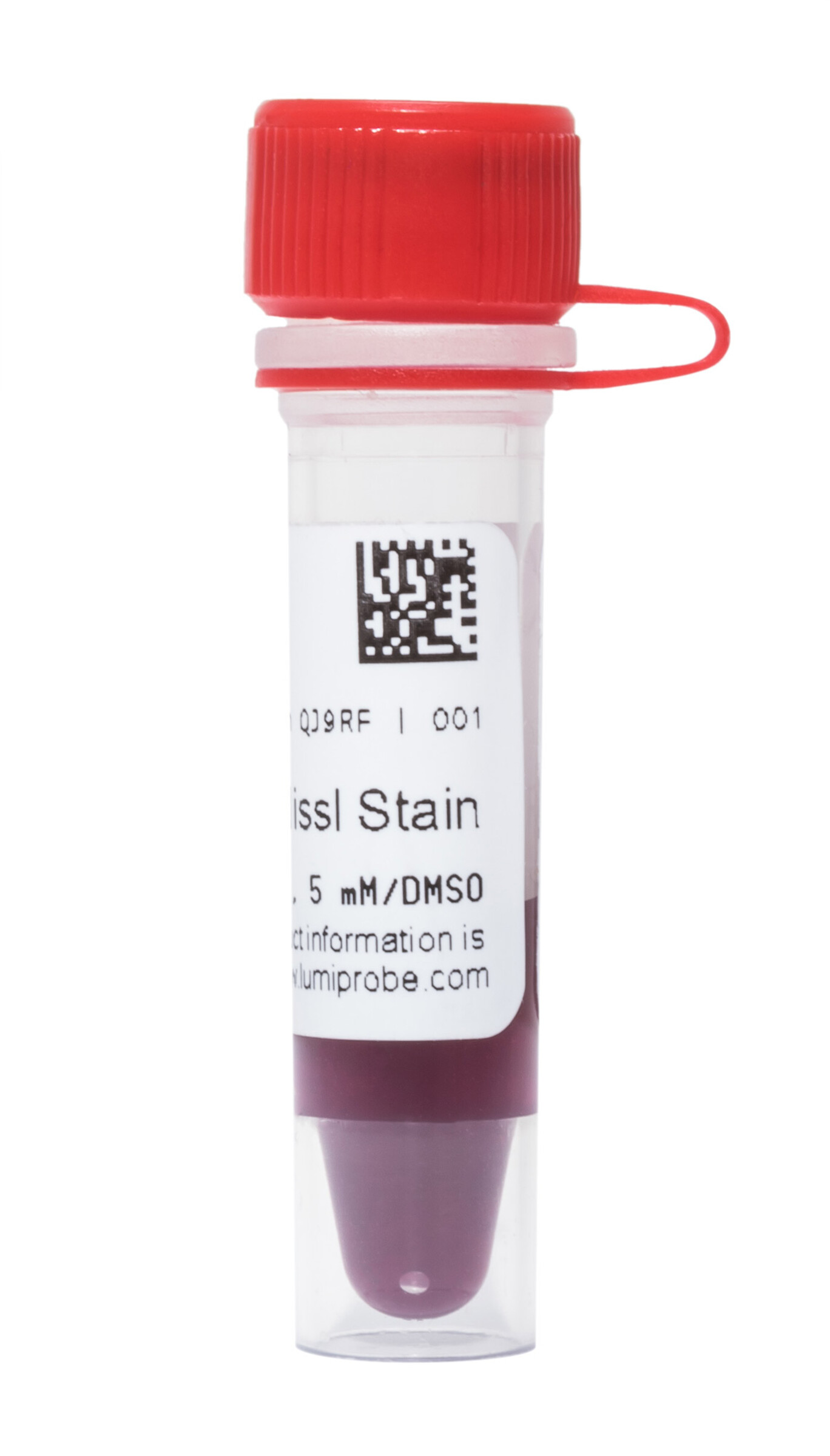
Red Fluorescent Nissl Stain
| Cat. # | Quantity | Price | Lead time | Buy this product |
|---|---|---|---|---|
| 1B010 | 50 uL |
–
|
in stock | |
| 2B010 | 500 uL |
$139.00
|
in stock |


Nissl staining is a widely used histological method for visualizing the morphology of nervous tissue. The method is based on the binding of basic dyes to cellular nucleic acids. Since the neuronal perikarya are characterized by intense protein synthesis and, consequently, a high content of ribosomal RNA in the rough endoplasmic reticulum (the so-called 'Nissl substance'), the neuronal cytoplasm stains significantly more intensely than their nuclei. This makes Nissl-stained neurons easily distinguishable from glial cells, making this method specific for neuron identification.
We offer highly concentrated (1,000×) Fluorescent Nissl Stains with different spectral properties.
Red Fluorescent Nissl Stain is a fluorescent dye that does not penetrate living cells and exhibits no fluorescence in the absence of nucleic acids. When bound to RNA and DNA, its fluorescence is greatly enhanced.
Red Fluorescent Nissl Stain is easily separated from the fluorescence of blue (CFP, DAPI, Hoechst), green (GFP, AF 488, FITC, LUTOX® Green), and far-red (AF 647, Cyanine5, 7-AAD, LDS 751) dyes, which allows it to be used for multi-color labeling of nervous tissue.


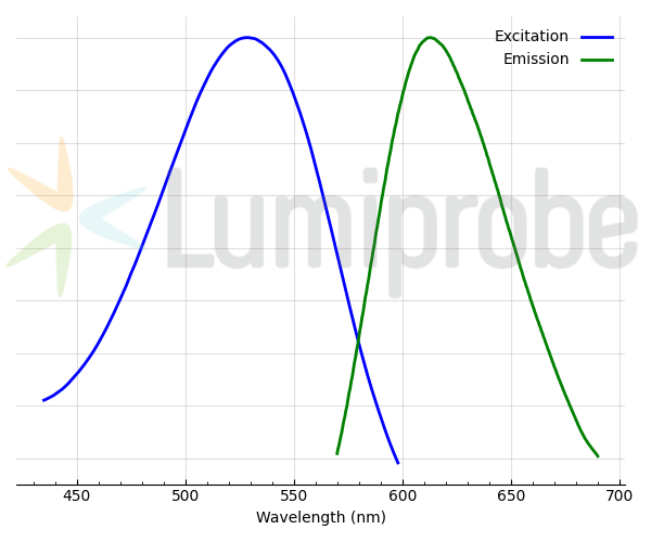
Excitation and emission spectra of Red Fluorescent Nissl Stain
Recommended protocol
Calculator
Customers also purchased with this product


General properties
| Appearance: | dark violet solution |
| Quality control: | NMR 1H and HPLC-MS (90+%) |
| Storage conditions: | 24 months after receival at -20°C in the dark. Transportation: at room temperature for up to 3 weeks. Desiccate. |
| MSDS: | Download |
| Product specifications |
Spectral properties
| Excitation/absorption maximum, nm: | 535 |
| Emission maximum, nm: | 613 |


 $
$ 
