The kits allow rapid introduction of fluorophores (fluorescent dyes) into an antibody (2-3 tags per 1 protein molecule). Each kit contains components for 10 reactions × 100 µg of antibody. The principle of action is based on using succinimide ester of the dye (taken in excess), which reacts with free amino groups of the antibody (N-terminal amino group, amino group on lysine) in a mild alkaline medium. Subsequent antibody purification from unreacted reagents is performed by gel filtration on microcolumns (included in the kit).
Kit components
| Kit component | Count | |||||||
|---|---|---|---|---|---|---|---|---|
|
1321-10rxn 10 reactions |
3321-10rxn 10 reactions |
7321-10rxn 10 reactions |
5321-10rxn 10 reactions |
6321-10rxn 10 reactions |
1821-10rxn 10 reactions |
6821-10rxn 10 reactions |
||
| N1320, sulfo-Cyanine3 NHS ester, 1 rxn | 10 | — | — | — | — | — | — | |
| N3320, sulfo-Cyanine5 NHS ester, 1 rxn | — | 10 | — | — | — | — | — | |
| N7320, sulfo-Cyanine5.5 NHS ester, 1 rxn | — | — | 10 | — | — | — | — | |
| N5320, sulfo-Cyanine7 NHS ester, 1 rxn | — | — | — | 10 | — | — | — | |
| N6320, sulfo-Cyanine7.5 NHS ester, 1 rxn | — | — | — | — | 10 | — | — | |
| N1820, AF 488 NHS ester, 1 rxn | — | — | — | — | — | 10 | — | |
| N2825, AF 594 NHS ester, 1 rxn | — | — | — | — | — | — | 10 | |
| A1115, Desalting spin column, PBS, 1 pcs | 10 | 10 | 10 | 10 | 10 | 10 | 10 | |
| Desalting receptacle vial, 1.5 mL | 10 | 10 | 10 | 10 | 10 | 10 | 10 | |
| Desalting waste vial, 2 mL | 10 | 10 | 10 | 10 | 10 | 10 | 10 | |
| PBS tablet, for 100 mL of buffer | 1 | 1 | 1 | 1 | 1 | 1 | 1 | |
| 15050, DMSO (dimethyl sulfoxide), labeling grade, 1 mL | 1 | 1 | 1 | 1 | 1 | 1 | 1 | |
| 1584-05mL, Sodium azide solution, 3%, 0.5 mL | 1 | 1 | 1 | 1 | 1 | 1 | 1 | |
| 1689-15mL, Sodium bicarbonate, 126 mg | 1 | 1 | 1 | 1 | 1 | 1 | 1 | |
Store at temperature between 4°C and 20°C. Do not freeze! Transportation: at room temperature for up to 3 weeks. Avoid prolonged exposure to light. Desiccate.
Shelf life 12 months.
Protocol
1. Antibody preparation
The antibody preparation must not contain impurities of free amino acids or other proteins, such as BSA, or components of buffer solutions with pH 2-7.5 or 9-12. If the antibody is in a pH 8-8.5 buffer (sodium bicarbonate or Tris-HCl) and is guaranteed to be free of other impurities, it can be used in the reaction without further purification. If you are unsure that the antibody preparation is impurities-free, perform a purification procedure. It is recommended to use one of the following methods for antibody purification (if necessary): dialysis, gel filtration, or ultrafiltration on columns, using 0.1 M sodium hydrogen carbonate solution*.
Optimal conditions for antibody labeling: 1 mg/mL antibody in 0.1 M sodium bicarbonate solution without impurities. The presence of the preserving agent sodium azide in the antibody solution (up to a concentration of 0.04%) does not affect the reaction.
If the antibody concentration is below 1 mg/mL, it must be brought to 1 mg/mL by concentration (ultrafiltration) and subsequent dilution. Two concentration/dilution cycles are recommended for complete purification from possible impurities. After each concentration procedure, the antibody is diluted with 0.1 M sodium bicarbonate. If the antibody concentration is higher than 1 mg/mL, washing the antibody once on an ultrafiltration column with 0.1 M sodium bicarbonate before dilution is recommended. Two concentration/dilution cycles are recommended to ensure complete purification from possible impurities. After washing, the antibody is diluted with 0.1 M sodium bicarbonate.
It is recommended to monitor the antibody concentration spectrophotometrically (a cuvette-free spectrophotometer is best suited for this purpose).
It should be noted that the antibody concentration in the reaction mixture affects the degree of labeling. For example, a reaction with 100 µg of antibody in a volume of less than 100 µL will yield a modified antibody with a labeling degree of up to 4-5 dye molecules per antibody. When the reaction is performed with 100 μg of antibody in a volume larger than 100 μL, a modified antibody with a labeling degree of 0.3-1 dye molecules per antibody will be obtained.
* To prepare 0.1 M sodium bicarbonate, add 15 mL of deionized water to the contents of the dry sodium bicarbonate tube included in the kit.
2. Setting up the reaction
2.1. Add a solution of the antibody in 0.1 M sodium bicarbonate to a test tube with a lyophilized fluorophore (fluorescent dye) (recommended amounts are 100 µg** antibody per 100 µL), vortex until the reagent is completely dissolved, and incubate for 30 min at room temperature.
** If it is necessary to react with a smaller amount of antibody, it is necessary to dilute the lyophilized reagent in 10 μL of anhydrous DMSO and take 1 μL of solution for the reaction for every 10 μg of antibody. The reagent solution in DMSO cannot be stored..
3. Purification of the labeled antibody
3.1 Dissolve the PBS buffer tablet in 100 mL of water.
3.2 Prepare the column. Make sure the column is at room temperature. Resuspend the gel on a vortex. Remove the cap from the column, place it in a waste receptacle, and centrifuge for 2 min at 1,000 g (the speed must be carefully controlled — for a standard 6 cm rotor, 1,000 g corresponds to 3,800 rpm). When centrifuging, the small bulge of the column should be oriented outwards. After centrifugation, remove the filtrate.
3.3 Apply 400 µL of PBS buffer to the column and centrifuge for 2 min at 1,000 g. Remove the filtrate after centrifugation. Transfer the column into the 1.5 mL antibody collection tube provided in the kit.
3.4 Apply 100 µL*** of reaction mixture onto the center of the column, incubate for 1 min, and elute the antibody by centrifugation for 2 min at 1,000 g. Purified antibody**** will collect in the tube.
*** For purification, 50 to 100 µL can be applied to the column. If the reaction was carried out in a smaller volume, it is recommended after the reaction to increase the volume of the preparation to at least 50 µL by adding PBS buffer.
**** After centrifugation, sodium azide solution (available in the kit) can be added to the antibody preparation in a volume of 1% of the preparation volume. If necessary, the antibody preparation can be divided into aliquots. The aliquot with which the work is carried out should be stored at 4 °C, and the remaining aliquots should be stored at −20 °C. Only solutions to which sodium azide has been added may be stored at 4 °C.
4. Measurement of dye to antibody ratio
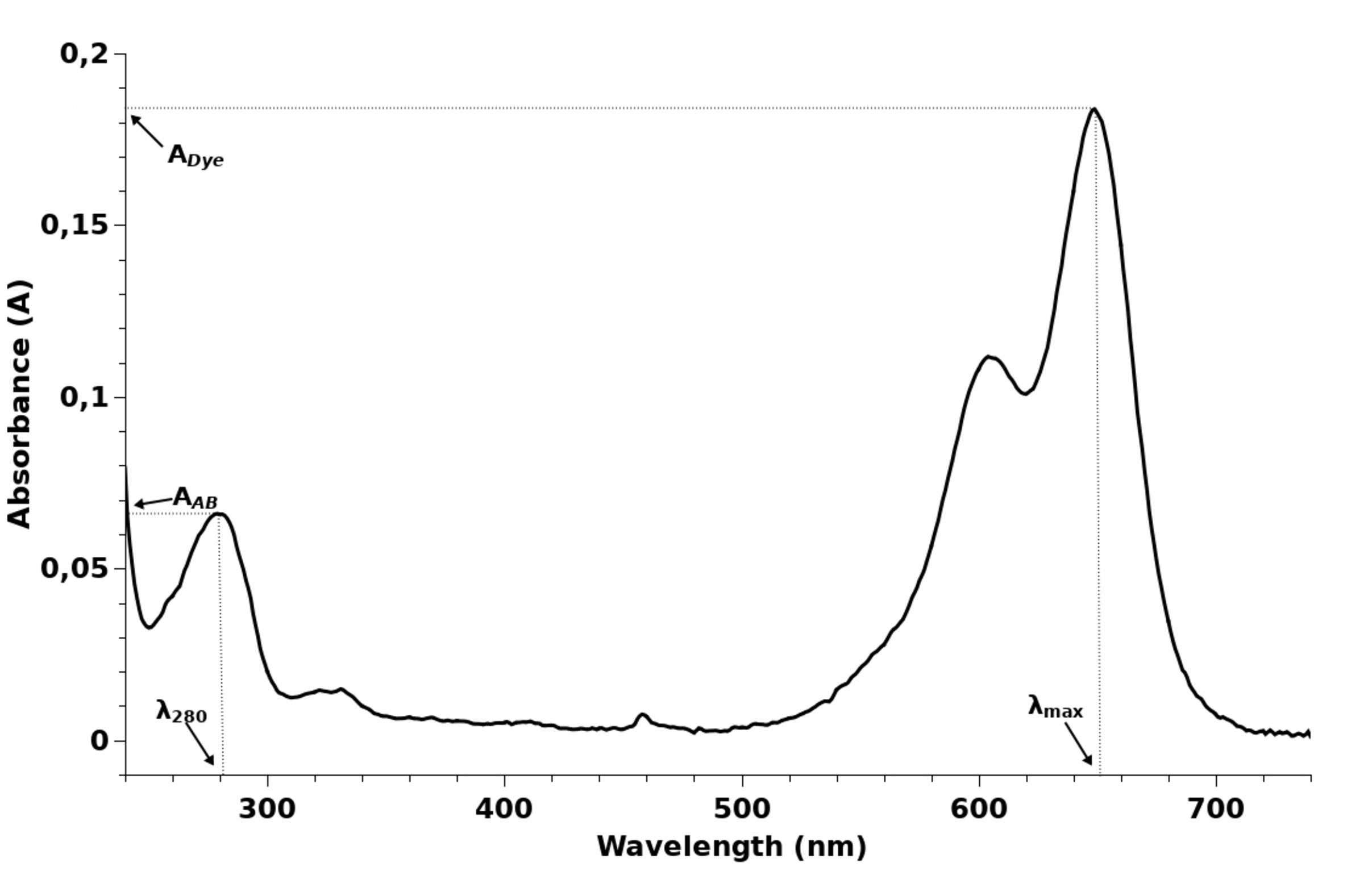
To calculate the dye-to-antibody ratio, measure the absorption spectrum of the conjugate, or absorption at 280 nm (AAB) and at dye absorption maximum (ADye). A typical absorption spectrum of a dye-labeled antibody is shown below. Depending on the dye, the wavelength of the maximum may vary.

An absorption spectrum of a labeled antibody contains a dye peak (with a longer wavelength) and an antibody peak (at around 280 nm). The dye-to-antibody ratio is calculated using the following formula:

where Dye/AB is the required number of dye molecules per antibody molecule, ADye is the optical density of the sample at the dye absorption maximum, AAB is the optical density of the sample at the wavelength of 280 nm, εAB is molar extinction coefficient of the antibody at 280 nm (210,000 for IgG), εDye is molar extinction coefficient of the dye at the wavelength of absorption maximum (taken from the table below), CF280 is correction factor for the dye at 280 nm (taken from the table below).
| Dye | λmax, nm | ε | CF280 |
|---|---|---|---|
| sulfo-Cyanine3 | 548 | 162,000 | 0.06 |
| sulfo-Cyanine5 | 646 | 271,000 | 0.04 |
| sulfo-Cyanine5.5 | 673 | 195,000 | 0.11 |
| sulfo-Cyanine7 | 750 | 240,600 | 0.04 |
| sulfo-Cyanine7.5 | 778 | 222,000 | 0.09 |
| AF 488 | 495 | 71,800 | 0.10 |
| AF 594 | 586 | 105,000 | 0.51 |
Example of calculation
After labeling of IgG antibody with sulfo-Cyanine5 and purification, a solution was obtained with absorption spectrum above. Determine dye-to-protein ratio.
Using the absorption spectrum, the following values can be found: ADye = 0.184 at dye absorption maximum (646 nm, see the table), AAB = 0.066 (at 280 nm), εAB = 210,000 for IgG; εDye = 271,000, and CF280 = 0.04.

Dye/AB — the number of dye molecules per one antibody molecule is 2.43.
Interpretation of results, and antibody storage
The optimal dye-to-antibody ratio is usually 2-3 dye molecules per antibody. Further increase of the dye loading does not improve the fluorescence signal as concentration quenching of fluorescence is observed. If the degree of labeling is insufficient, the amount of antibody introduced into the reaction should be reduced. Low labeling may be due to the use of an expired kit. We recommend to test binding of labeled antibodies with their antigens.
The labeled antibodies can be stored at −20 °С. An aliquot currently in use should be stored at 4 °С to avoid repeated freezing-thawing. The stability of the conjugate is determined by the antibody itself rather than the fluorophore or the chemical bond. Labeled antibodies should not be exposed to direct sunlight for an extended time but are quite compatible with ambient light.
Related kits
AF 488 antibody labeling kit
Ready-to-use kit for labeling antibodies and other proteins with AF 488 dye via NHS ester chemistry. The kit contains all necessary chemicals and consumables.| Cat. # | Quantity | Price | Lead time | Buy this product |
|---|---|---|---|---|
| 1821-1rxn |
1 rxn
|
$69
|
1 days | |
| 1821-10rxn |
10 rxn
|
$450
|
1 days |

AF 594 antibody labeling kit
Ready-to-use kit for labeling antibodies and other proteins with AF 594 dye via NHS ester chemistry. The kit contains all necessary chemicals and consumables.| Cat. # | Quantity | Price | Lead time | Buy this product |
|---|---|---|---|---|
| 6821-1rxn |
1 rxn
|
$69
|
1 days | |
| 6821-10rxn |
10 rxn
|
$450
|
1 days |

sulfo-Cyanine3 antibody labeling kit
Ready to use kit for the labeling of antibodies and other proteins with sulfo-Cyanine3 dye via NHS ester chemistry. The kit contains all necessary chemicals and consumables.| Cat. # | Quantity | Price | Lead time | Buy this product |
|---|---|---|---|---|
| 1321-1rxn |
1 rxn
|
$69
|
1 days | |
| 1321-10rxn |
10 rxn
|
$450
|
1 days |

sulfo-Cyanine5.5 antibody labeling kit
Ready-to-use kit for labeling antibodies and other proteins with sulfo-Cyanine5.5 dye via NHS ester chemistry. The kit contains all necessary chemicals and consumables.| Cat. # | Quantity | Price | Lead time | Buy this product |
|---|---|---|---|---|
| 7321-1rxn |
1 rxn
|
$69
|
1 days | |
| 7321-10rxn |
10 rxn
|
$450
|
1 days |

sulfo-Cyanine5 antibody labeling kit
Ready-to-use kit for labeling antibodies and other proteins with sulfo-Cyanine5 dye via NHS ester chemistry. The kit contains all necessary chemicals and consumables.| Cat. # | Quantity | Price | Lead time | Buy this product |
|---|---|---|---|---|
| 3321-1rxn |
1 rxn
|
$69
|
1 days | |
| 3321-10rxn |
10 rxn
|
$397
|
1 days |

sulfo-Cyanine7.5 antibody labeling kit
Ready-to-use kit for labeling antibodies and other proteins with sulfo-Cyanine7.5 dye via NHS ester chemistry. The kit contains all necessary chemicals and consumables.| Cat. # | Quantity | Price | Lead time | Buy this product |
|---|---|---|---|---|
| 6321-1rxn |
1 rxn
|
$69
|
21 days | |
| 6321-10rxn |
10 rxn
|
$450
|
21 days |

sulfo-Cyanine7 antibody labeling kit
Ready-to-use kit for labeling antibodies and other proteins with sulfo-Cyanine7 dye via NHS ester chemistry. The kit contains all necessary chemicals and consumables.| Cat. # | Quantity | Price | Lead time | Buy this product |
|---|---|---|---|---|
| 5321-1rxn |
1 rxn
|
$69
|
1 days | |
| 5321-10rxn |
10 rxn
|
$450
|
1 days |


 $
$ 

